- Write a project abstract : Pick up something you heared this week. Think about a project and write a “grant proposal” kind of thing. Send the final version by Monday. [deadline]
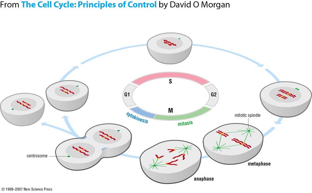
Introduction to the Cell Cycle
the DNA molecule: Long-term storage of information
- reading
- transmission
The human karyotype: 23 chromosome pairs
During cell division, you seperate the chromatins, not the chromosomes.
The human body experiences ~10, 000 trillion cell divisions during the lifetime.
Why do cells divide?
- Reproduction: the only time when the chromosome pairs reseperate
- Regeneration: Starfishes are really good at it, Human liver can regenerate from its $1/3^{rd}$ proportion.
- Cell Rejuvination

Most cells are usually in the $G_0$ phase.
The “must do” of cell division:
- replicate their DNA and organelles
- segregate its contenet equally in the two daughter cells
- ensure the right order of events
Cell mass grows linearly during the cell cycle, whereas, the DNA content looks like a step function.
The search for the master cell cycle regulators:
- Biochemical approach: Cells specialized for rapid and synchronomus division (exhibiying natural blocks)
- Genetic approach: Cells in whiuch genetic manipulations are straightforward. Cells displaying prominent control points.
- Eventuall, the two branches were merged.
A temperature sensitive mutant: The mutant will die due to temperature change. Its a way of implementing conditional muatation in the experiment cycle.
Haploid yeast: Its easier to screen for mutants.
Permissive (low) temperature: You see all stages of the cell cycle.
Restrict (high) temperature: Cells are stuck at the $G_1$ phase.
Cyclin + Cdk complex are essential for cell division in frog egg. The 2001 Nobel Prize for given for the discovery of key regulators of the cell division cycle.
Regulators of the cell cycle:
- G1: Cyclin D + Cdk 4 and Cyclin D + Cdk6
- S. Cyclin E + Cdk2
- G2: Cyclin A + Cdk2
- M: Cyclin B + Cdk1
Whwn something decreases very sharply, its probaly being regulated by the degradation os something.
DNA replication “inside a cell”
There are restrictions when we need cell replication inside the cell, in contrast to say a PCR:
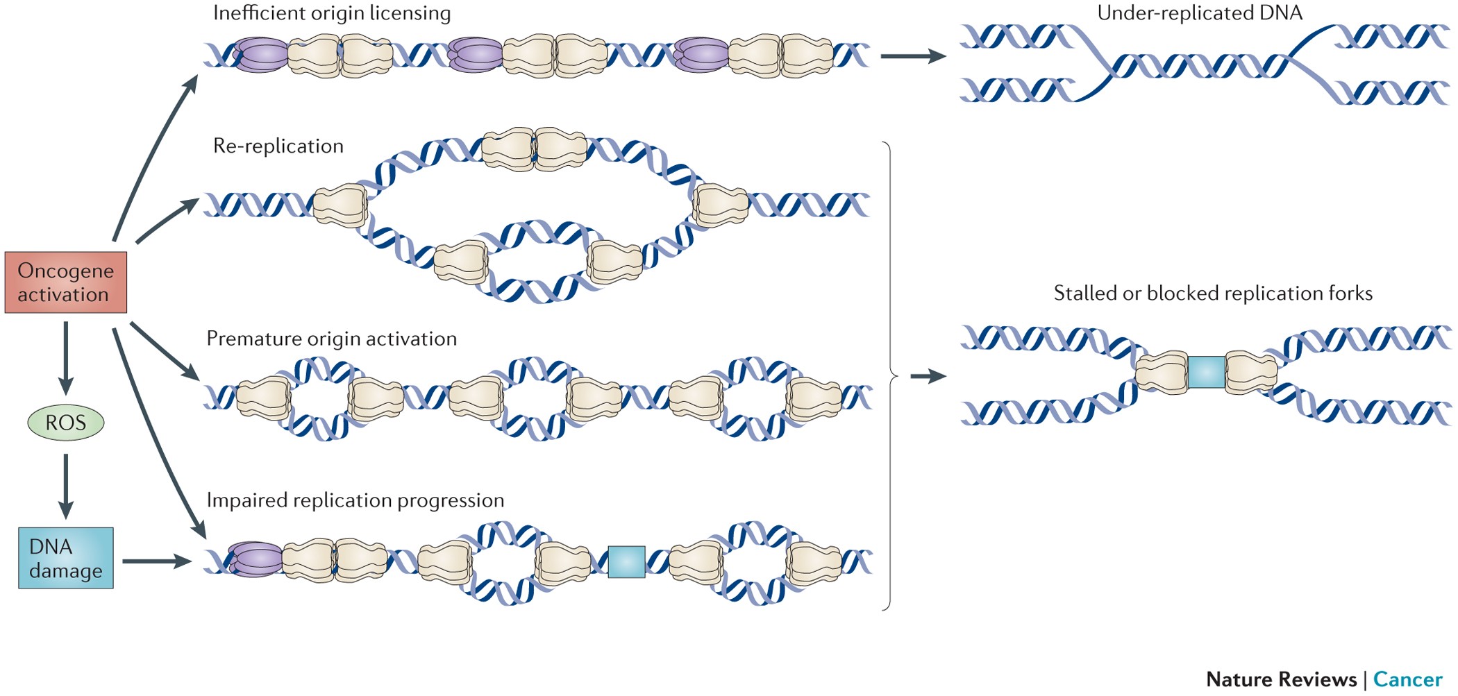
Replication is done with only one cycle.
Under-replication / Over-replication of the DNA can lead to misfolding of the chromatins.

In bacteria, the DNA replication is not very time controlled, hence the problem of over-replication does not hinder the development of the cell.
Origin is liciened to “ORC” : Origin Recognition Complex, which recognises the Helicase.
Helicase: Enzyme that unpacks the genetic material. In this context, it opens up the double Helix.
What makes certain Origins slower? (Some origins can fire but they don’t)
To prevent the activation of certain Origins, the cell can use checkpoints like the regulation of DDK. mec1 modification can express DDK and r+then control the oroduction of DDK.
Mitosis
Riddle: Two blind man go shopping for socks
There was a man who went to the mall and he bought 3 pairs of red socks and 3 pairs of white socks. Another man who already bought 3 pairs of red socks and 3 pairs of white socks came back to return his 3 pairs of red socks and 3 pairs of white socks. They are both blind. As they were walking they bumped into each other. All the socks scattered around the floor, but each pair remained held together by a rubber band. Nobody helped them pick it up except each other, but in 3 minutes they both put them back altogether. Each man ended up with the same colors of socks he started with: six red and six white. How is that possible if they are blind?
How do cells divide?
Kangaroo-Rat from Australia as our model organiosm, since it has only 12 to 13 chromosomes.
Insects (like the Drosophilia) divide for 13 generations in a single “Pangea cell”. Hence, the division is extremenly synchronised.
Questions
The constraint of space. Can’t we have a bigger nucleus? So that more space allows for less crowding bias.
How much force does the spindles apply when they are pulling apart the chromatins?
How do they [the spindles] know when its the right time to pull ?
How do Chromosomes find their way?
Several models have been proposed. Mathematically, it was found that a random microtuble search of the space would not be enough.
The number of mechanisms that promote effcient chromosome capture are about 6-7 in number and still increasding.
Answers
The correct position to grab the chromosome is done using mechanosensing. A tension-sensitive error correction mechanism is used.

Merotelic attachment is a problematic case and is responsible for most errors during copying.
Metaphase $\rightarrow$ Anaphase : Separase is activated by the APC. It is the primary regulatory mechanism.
The seperation mechanism relies on sensing all microtubles attachment to kinetochores.
~700 pN force is applied by the spindle.
Cell Division in Developement
Cell Polarity: Eg: Neurons; Edourard Van Beneden and Carl Rabl.
~75% of human diseases have analogues/homologues in Drosophila.
“They don’t eat much, reproduce like mad, and never demand an ethics committee” - whyfiles.org
PAR-genes controls the cell polarity. The spatial clue is the Sperm centrosome.
Making polarized cells:
flowchart TD Spatial-Cue--Symmetry-breaking-->2(Distribution of Polarity determinants) 2 --> XCell Polarity is observed in multiple cell contexts.
Why do we care about epithelial organisation? Epithelial barriers are essential for organism compartmentalization.
“Apical-basal”: “inside and outside” maybe
Epithelial cell devision in the basal part is spatially oriented. We see Planar spindle orientation that precedes planar cell division.
Epithelial cell division:
- Asymmetric division: Different fates/ Tissue thickening
- Symmetric division: Identical fates/Tissue spreading
The Position of the centrosomes determine the positioning of the spindle orientation.
Hertwig “long-axis” rule states that cells prefer to divide perpendicular to the long axis of the mitotic spindle.
Polarity oscillations maintains tissue architecture.
Tublin Cytoskeleton/ Centrosomes (general principles)
All functions of the cell kind of depend on the cytoskeleton.
Tublins: are the functional building blocks.
Filament polarity largely determines the complex structure of cells and its polaritty.
Microtubles direct vesicle trafficked by acting as tracks for motor proteins. Golgi and lysosomes move towards minus ends while secretory vescicles move towards at the plus end.
You have motor proteins moving across this polar track:
- Dyneins:
- Kinesins:
Dybamic Instability is driven by GTP hydrolysis. GTP units like to be straight but the GDP units like to be curved.
Cellular factors regulate Dynamc Instability: Cyclines and Cdks also change the structure of the cytoskeleton dynamics.
Aggregation follows bulk polymerization dynamics.
The cell controls where polymers from using nucleating factors.
Centrosomes contains $\gamma$-tublin which acts as nucleating sites for Tublin filamentation.
Henneguy-Lenhossek hypothesis: Centrosomes can form spindles as well as flagella. They also form cilia.
Most human cells have cellia, hence, problems with cillia results in a myriad of seemingly unconnected pathologies. Like Retinal degeneration, Kidney Cysts, Cognitive defects.
Cillia Assembly: The centrosomes goes to the membrane and starts assembling a microtuble structure called the Cillium.
Centrioles are not necessary for cell division. Cell divisions can happen without it. Many organism families have dropped it altogether.
A centrosome has two centrioles.
Chromosomes can also nucleate microtubles, hence, centrosoles are redundant for cell division.
Human/Mice Cells need Centrioles for cell division.
Kinesisns can also bind together to focus microtuble spindles.
More than one centrosome can cause a variety of cancers because they can interfere during cell division and cause aneuploidy.
Cells adapt to multiple centrosomes by clustering them, especially cancer cells.
Senior IGC Fellow: Pedro
Bacteria have something similar to Actin and Tubilin.
Cancer Centrosomes & Cancer Mitosis
Asymetric cell division
Aneuploidy
Increased/Decreased signalling plateform
Increased Microtuble directed polarization
Altered Rho GTPhase activity andd Altered Focal adhesion turnover
Membrane Protein Proteostasis
30% of proteins encoded are pushed into the Endoplasmic Reticulum, and are membrane or secretary proteins.
We have organelles to sepertate many different chemical processes : to compartmentalize them.
Fluid Mosaic Model:1972 to present

Transmembrane domains are alpha helices that can be used to classify memebrane proteins
Polytopic membrane: TODO
Charged residues are used to emable polymerization or dimerisation.
~50% of membrane is composed of transmembrane proteins.
Membrane proteins are often difficult to fold. And a few disease causing mutations can cause a variety of debilitating diseases.
The ER is responsible for the quality control of proteins. Folding is tighly linked to the oscialltions of proteins. Addition of sugars.
Ramanujan S. Hegde for Membrane Protein studies.
The signal sequence hypothesis: As the polypeptide emerges from the ribosome, it is sensed and inserted in the membrane as it folds.
Signal peptide or transmembrane domains are needed to recognize a transmembrane protein. Tail Anchored (TA) proteins have a very short C terminal and no signallling domain, hence co-translation is not possible.
Post-translation; TODO
Lipid Droplets: Intracellular stores of triglycerides & cholestryl esters
- Organalles: They have their own specialised proteiome.
- Features & contents of liquid droplets: Phospholipid Monolayer and TAGs and Sterol ethers in the inside.
- Their function is to store energy in the form of Tri-glycerides.
- viral assembly plateforms
- inflammatory lipid biosynthesis
- TODO
- ERAD: ER-associated degradation for misfolded proteins.
New Paradigms in Viruses
Viruses infects every organism. For example, Hepatitis B is prevelent among a lot of organismic families.
Most epidemics and pandemics have been caused by viruses.
Its very diffiult to have animal models where you can study viruses generally.
If they don’t find a receptor, they are innocuous.
Host spillover events: virus finds a new host while jumping from its current “natural” host.
Viruses modify the Cell architecture/pathways/homeostasis.
Viral lifecycle:
flowchart LR 1(x) --> 2(x) 2 --> 3(x) 3 --> 4(x)Influenza A virion: a small virus: ~13kB
Correct grouping of the RNA segments is necessary for viral replication. For example in Influenza A, there are 8 RNA segments.
Viral inclusion: It is the seperation of viral components in the cytoplasm mediated by liquid-liquid phase seperation or !percolating! loose RNA-RNA interactions.
IAV inclusions have the properties of liquid:
- Fusion and division
- Internal rearrangement
- Fast diffusion
- Dissolve quickly (responds to stimuli)
Liquid-Liquid Phase Seperation in Biology
Liquid-Liquid phase seperation:
Brangwynne et alMembraneless organelles behave like liquid droplets.
Progress with Live Cell Imaging has been a priminent reason for the discovery in Liquid-Liquid Phase seperation.
First diffraction patterns were derived from viruses because of their symetrical structures. And they are easier to crystallize viruses.
Crystallization was used as a purification technique for viruses and protein for years.
There has been a lot of progress in Live Cell imaging without tags (Differential/Nomarski interference contrast microscopy - DIC; Holographic Interference Microscopy - HIM etc)
Ethanol is added during protein precepitation with salts to change the dielectric constant of the solvent.
Types of Interactions, for species $a, b$:
- Homotypic interaction energies: $E_{aa}, E_{bb}$
- Heterotypic interactions: $E_{ab}$
For L-L pahse separation: $$ \chi = \frac{E_{heterotypic} - E_{homotypic}}{k_BT} » 1 $$
Valency of interactions:
- Avidity: Multiple distinct pairwise interactions among binding sites and receptors
- Allovalency: Multiple epitopes along the same legands.
“Fuzzy interactions” : Multiple interchangable interaction sites.
At concentrations above $C_{critical}$, a protein will form droplets.
Limiting Factor for an infinite protein droplet radius:
- Conncentration outsiede the droplet ($C_{out}$)
- Temperature fluctuations
Zeta potential and the surface charge
Biomolecular Condensate is a mix of ‘Scaffolds’ and their ‘Clients’ RNAs can also act as ‘Scaffolds’.
Ligand binding might be necessary for IDFs to go from a disordered state to an ordered transition.
Some residues are prevalent in proteins that undergo LLPS.
Coacervates: TODO
Simple condensates like to exclude everything within them. So they generally have no aqueous pockets/capsules inside them.
